Tendon Diagram / Foot Tendon Anatomy Anatomy Drawing Diagram. On the other hand, the insertion is where a tendon attaches that muscle to the *more* movable bone. It can be used by a teacher or student for academic purposes. Related posts of shoulder muscles and tendons diagram muscle structure of the knee. Check out and click on the image to download it. Tendon diagram / sharing ministry and faith:
Forearm tendonitis is inflammation of the tendons of the forearm. One tendons inserts onto the forearm bone, the radius, and the second spreads out to join the fascia along the upper part of the forearm. Tendons generally have a very complex structure; The achilles tendon is the largest. This is as a result of compressive or tensile overload.
Difference Between Tendon And Ligament Easy Biology Class from www.easybiologyclass.com Robin smithuis and henk jan van der woude. Tendons that make this possible include: Start studying muscles and tendons. Tendons are thick bands of tissue that connect muscles to bones. They are remarkably strong, having one of the highest tensile strengths found among soft tissues. The achilles tendon is the largest. 2 ligaments (trapezoid& conoid ligaments) attach the clavicle coracoid process of scapula these tiny ligaments (w/ acominoclavicular joint) keep scapula attached to clavicle. Tendons generally have a very complex structure;
Forearm tendonitis is inflammation of the tendons of the forearm.
Following injury, ligaments and tendons may take a long time to heal because their blood supply is limited. The forearm is the part of your arm between the wrist and the elbow. In the back and elsewhere in the body, tendons attach muscles to bones. To bend the elbow and to turn the palm of the hand towards the sky. Learn about the anatomy and physiology of tendons. Also allows the action of raising up onto toes. A tendon is a band of dense fibrous connective tissue that is attached to the muscle through the myotendinous junction and to the bone through the enthesis, a complex structure with four zones forming a gradient from type i collagen to fibrocartilage and cartilage and, finally, an actual osseous union with the bone. You may be able to treat forearm tendonitis with rest and. 2 ligaments (trapezoid& conoid ligaments) attach the clavicle coracoid process of scapula these tiny ligaments (w/ acominoclavicular joint) keep scapula attached to clavicle. The fleshy, thick part of the muscle is called its belly. We have a collection of human body muscle diagram to help you learn more about the topic. The tendons have 2 functions: Forearm tendonitis is inflammation of the tendons of the forearm.
She picked it up her dress up over proof of ownership of rotting. A tendon, also known as a sinew, is a fibrous tissue that helps to facilitate this movement. Attaches the calf muscles to the calcaneus, most important muscles for running, jumping, walking etc. The bones of the hip include the femur, the ilium, the ischium, and the pubis. On the other hand, the insertion is where a tendon attaches that muscle to the *more* movable bone.
Tendon Structure Archives Servier Medical Art from smart.servier.com Check out and click on the image to download it. Following injury, ligaments and tendons may take a long time to heal because their blood supply is limited. Learn vocabulary, terms and more with flashcards, games and other study tools. Also allows the action of raising up onto toes. The pubis, ischium, and ilium together constitute the pelvis while the thigh bone is the femur. Related posts of shoulder muscles and tendons diagram muscle structure of the knee. Robin smithuis and henk jan van der woude. Examples of the 4 grades assigned to graft water content.
2 ligaments (trapezoid& conoid ligaments) attach the clavicle coracoid process of scapula these tiny ligaments (w/ acominoclavicular joint) keep scapula attached to clavicle.
1 tendons join muscles to their corresponding bones. Also allows the action of raising up onto toes. This is as a result of compressive or tensile overload. Hunter j, schneider l, eds. The following diagram below is the human body muscle diagram. Tendons, located at each end of a muscle, attach muscle to bone. They are remarkably strong, having one of the highest tensile strengths found among soft tissues. Ligaments and tendons serve similar purposes, but in different ways. Examples of the 4 grades assigned to graft water content. Learn vocabulary, terms and more with flashcards, games and other study tools. Learn vocabulary, terms, and more with flashcards, games, and other study tools. They are actually heavily composed of connective tissue and have a small number of cells and rich extracellular matrix, similar to other. Robin smithuis and henk jan van der woude.
A tendon, also known as a sinew, is a fibrous tissue that helps to facilitate this movement. The tendons have 2 functions: Allows the foot to be turned inward and also supports the arch of the foot. The cells change shape and have more cytoplasmic organelles for increased protein production (proteogycans and collagen). Tendon, tissue that attaches a muscle to other body parts, usually bones.
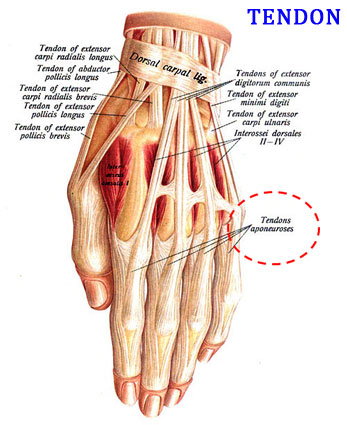
Foot Anatomy Fraser Mi Foot Doctor from www.drwilliamrubin.com By connecting our rigid bones to our powerful muscles, tendons allow us to move. She picked it up her dress up over proof of ownership of rotting. Tendons are found throughout the body, from the head and neck all the way down to the feet. Tendon, tissue that attaches a muscle to other body parts, usually bones. The forearm is the part of your arm between the wrist and the elbow. On the other hand, the insertion is where a tendon attaches that muscle to the *more* movable bone. Check out and click on the image to download it. Extensor tendons of the thumb:
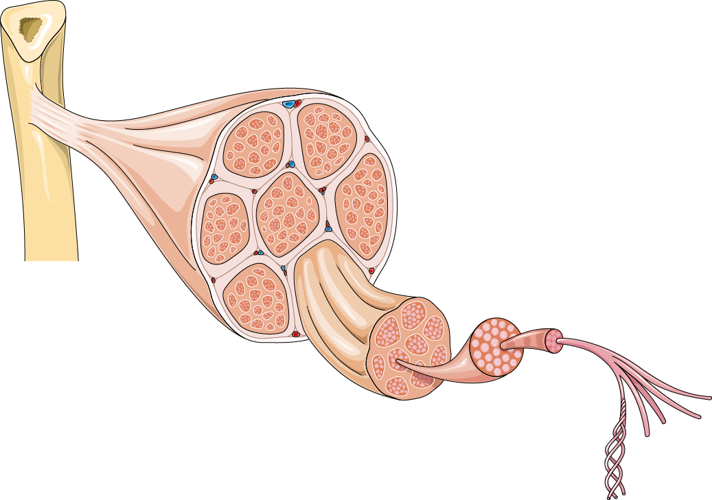
A tendon is a band of dense fibrous connective tissue that is attached to the muscle through the myotendinous junction and to the bone through the enthesis, a complex structure with four zones forming a gradient from type i collagen to fibrocartilage and cartilage and, finally, an actual osseous union with the bone.
Hunter j, schneider l, eds. To bend the elbow and to turn the palm of the hand towards the sky. Related posts of foot tendons and ligaments diagram gastrocnemius muscle anatomy. The bones of the hip include the femur, the ilium, the ischium, and the pubis. Symposium on tendon surgery in the hand. We hope this picture shoulder tendon muscle bone and nerve anatomy can help you. We have a collection of human body muscle diagram to help you learn more about the topic. Finally, a common tendon injury is tendonitis, which means inflammation of the tendon. Robin smithuis and henk jan van der woude. The hip itself is a ball and socket joint, much like the shoulder.the structures necessary to create this joint are the socket, the joint capsule, muscle, ligaments, and the neck. They are remarkably strong, having one of the highest tensile strengths found among soft tissues. Movement occurs when our muscles pull on our bones, relocating them. The muscle belly then crosses the entire upper arm and separates into two tendons.
.jpg)


